Jump to...
- Cerebrospinal fluid
- Reasons why you might need a CSF MCS
- How is the test done?
- What is a lumbar puncture?
- What are the risks associated with a lumbar puncture?
- How should you prepare for a lumbar puncture?
- What should you expect during a lumbar puncture?
- Laboratory testing and analysis of CSF
- Interpreting your test results
- Conclusion
Cerebrospinal fluid (CSF) analysis is a collective term for CSF microscopy, culture, and sensitivity.
CSF analysis is a way of looking for conditions like bacterial, viral, parasitic, or fungal infections that affect your central nervous system (brain and spine).
It can also be used to detect various autoimmune disorders, such as the presence of any primary tumor present in the central nervous system or its spread to the central nervous system (known as metastatic cancer).
It’s a series of laboratory tests performed on a sample of CSF.
The test mainly confirms central nervous system (CNS) infection not only before the treatment but also during and after the treatment procedure.
Cerebrospinal fluid (CSF)
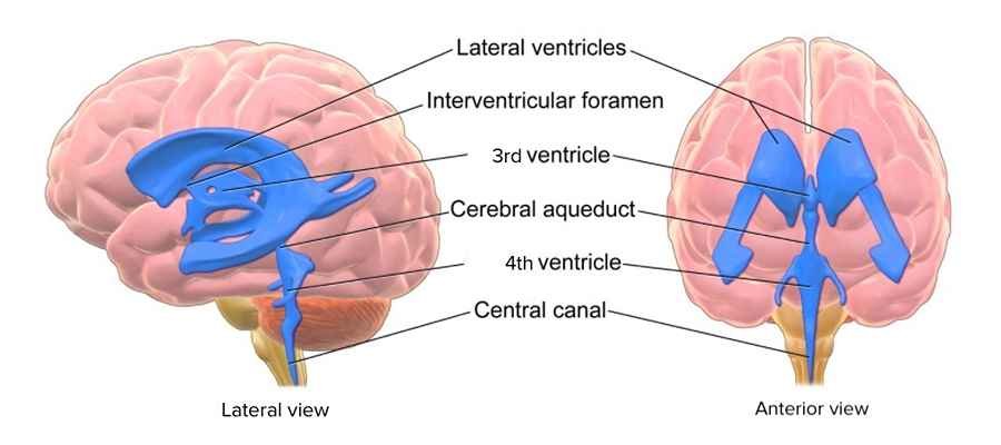
CSF is the clear fluid that cushions and delivers nutrients to your central nervous system (CNS).
The CNS consists of the brain and spinal cord.
CSF is produced in the brain and then reabsorbed into your bloodstream.
The fluid is completely replaced every few hours.
In addition to delivering nutrients, CSF flows around your brain and spinal column, providing protection and carrying away waste.
Reasons why you might need the test
A CSF analysis may be ordered if you’ve had CNS trauma.
It may also be used if you have cancer and your doctor wants to see if the cancer has spread to the CNS.
In addition, a CSF analysis may be ordered if you have one or more of the following symptoms:
- severe, unremitting headache
- stiff neck
- hallucinations, confusion, or dementia
- seizures
- flu-like symptoms that persist or intensify
- fatigue, lethargy, or muscle weakness
- changes in consciousness
- severe nausea
- fever or rash
- light sensitivity
- numbness or tremor
- dizziness
- speaking difficulties
- trouble walking or poor coordination
- severe mood swings
- intractable clinical depression
How is the test done?
The primary step in a CSF analysis is sample collection, which is your CSF.
It is collected through the process of lumbar puncture.
What is a lumbar puncture?
A lumbar puncture is sometimes called a “spinal tap.”
It’s a medical procedure that can involve collecting a sample of cerebrospinal fluid (CSF).
What are the risks associated with a lumbar puncture?
A lumbar puncture is generally considered safe, but it can involve some risks.
According to the Mayo Clinic, up to a quarter of people who get a lumbar puncture develop a headache afterward.
Lying down for a few hours after the procedure may lower your risk of getting a headache.
Other potential risks include tenderness or pain in your lower back and bleeding near the puncture site.
You may experience some pain and numbness that shoots down your legs.
In rare cases, people experience brainstem herniation, which is the movement of brain tissue from its normal position in the skull.
This is uncommon.
How should you prepare for a lumbar puncture?
Tell your doctor about all of the medications you’re taking, and ask them if you should stop taking any of them before your lumbar puncture.
For example, they may advise you to stop taking blood thinners, such as aspirin or warfarin.
Your doctor may also order a CT or MRI scan before your lumbar puncture.
They can use it to check for signs of swelling around your brain or other problems.
What should you expect during a lumbar puncture?
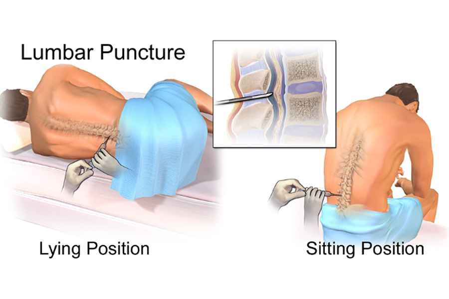
A lumbar puncture generally takes less than 30 minutes.
It’s performed by a doctor who is specially trained to collect CSF.
CSF is usually taken from your lower back area or the lumbar spine.
It’s very important to remain completely still during the procedure, as this way you avoid incorrect needle placement or trauma to your spine.
- You may be seated and asked to lean over so that your spine is curled forward. Or your doctor may have you lie on your side with your spine curved and your knees drawn up to your chest.
- Curving your spine makes a space between your bones in the lower back.
- Once you’re in position, your back is cleaned with a sterile solution. Iodine is often used for cleaning.
- A sterile area is maintained throughout the procedure. This reduces the risk of infection.
- A numbing cream or spray is applied to your skin.
- Your doctor then injects an anesthetic. Once the site is fully numb, your doctor inserts a thin spinal needle between two vertebrae.
- A special type of X-ray called fluoroscopy is sometimes used to guide the needle.
- First, the pressure inside the skull is measured using a manometer. Both high and low CSF pressure can be signs of certain conditions.
- Fluid samples are then taken through the needle. When the fluid collection is complete, the needle is removed.
- The puncture site is cleaned again, and a bandage is applied.
- You’ll be asked to remain and lie down for about an hour. This reduces the risk of a headache, which is a common side effect of the procedure.
Laboratory testing and analysis of CSF
The following are often measured in CSF analysis:
- white and red blood cell count (Microscopy)
- chloride
- glucose, or blood sugar
- glutamine
- lactate dehydrogenase, which is a blood enzyme
- antigens, or harmful substances produced by invading microorganisms
- total proteins
- oligoclonal bands, which are specific proteins
- cancer cells
- viral DNA
- antibodies against viruses (Sensitivity)
- bacteria (culture); they check to detect the state of the germs, i.e., when germs cause infection to grow, they identify the culture as positive, while in cases of no growth in germs, they conclude the culture is a negative test. Pathologists use microscopes or various chemical tests to identify the germ. Bacteria often grow within only 2 days, while others, including fungi, may take relatively more time to grow in the human body.
Interpreting your test results
Normal results mean that nothing abnormal was found in the spinal fluid.
All measured levels of CSF components were found to be within the normal range.
Abnormal results may be caused by one of the following:
- A tumour
- Metastatic cancer
- Hemorrhage
- Encephalitis: An inflammation of the brain
- An infection
- An inflammation
- Reye’s syndrome, which is a rare, often fatal disease affecting children that is associated with viral infections and aspirin ingestion,
- Meningitis, which you can get from fungi, tuberculosis, viruses, or bacteria
- Viruses such as West Nile or Eastern equine
- Guillain-Barré syndrome: is an autoimmune condition that causes paralysis and occurs after viral exposure
- Sarcoidosis, which is a granulomatous condition of unknown cause affecting many organs (primarily the lungs, joints, and skin),
- Neurosyphilis: happens when an infection with syphilis involves your brain
- Multiple sclerosis: is an autoimmune disorder that affects your brain and spinal cord
Conclusion
CSF microscopy, culture, and sensitivity tests have found wide usage in the diagnosis of many health conditions and diseases that affect the central nervous system.
These may be of four different categories: infectious diseases, which may be caused by viruses, bacteria, parasites, and fungi, while separating them from any other health condition.
I hope this piece helped.
You can leave your comments below, and I will reply to them.