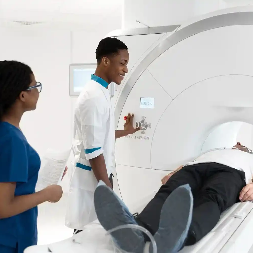
An X-ray is the most commonly performed imaging procedure in the hospital.
It is a brief, painless, safe, and non-invasive procedure that involves directing a small dose of a controlled beam of ionizing radiation called X-rays into the human body to take a picture of internal structures for a medical purpose.
A chest X-ray involves directing this beam of X-rays to the chest, thus providing images of the airway, lungs, heart, blood vessels, and bones.
Your doctor can detect abnormalities in these tissues or may see abnormal collections of water, pus, or blood within the lungs or surrounding the heart, or an abnormal collection of water, pus, blood, or air in the space surrounding your lungs.
Possible reasons to request a chest X-ray
Your doctor may request a chest X-ray for a wide range of reasons.
In emergencies, it may show injuries to chest structures that may result from a blunt or penetrating injury to the chest.
Some symptoms may warrant your doctor’s request for a chest X-ray, such as:
- Prolonged and/or severe cough
- Chest pain
- Breathing difficulties.
- Pre-employment medical fitness, Immigration screening,
- Position medical equipment in the chest to make sure it is in the right place. It can also be used in situations where an implanted medical device is displaced from the appropriate position, such as a chest tube, nasogastric tube, or endotracheal tube.
- To monitor the progress of treatment, e.g., in patients taking medication for tuberculosis.
What should you do before the scan?
The chest X-ray does not require any special preparation.
Usually, you would be scheduled on the day and time that the procedure would take place, which may last about 15-20 minutes from start to finish.
In the case of an emergency chest X-ray, you may be wheeled from the emergency department into the X-ray unit or room.
Pieces of jewelry, sunglasses, and body piercings will have to be taken off.
If you have any implanted object, either a medical device, such as a cardiac pacemaker, or nonmedical, such as jewelry, that can’t be taken off, please inform your doctor.
These objects can appear in the final picture as artifacts and may affect the reading and interpretation of the X-ray findings.
Also, remember to inform your doctor if you are pregnant or think you may be pregnant, as the procedure may differ.
This is to prevent any harm to the developing baby that can occur following exposure to ionizing radiation.
Be sure to wear clothing that can easily be removed and worn because the procedure would require you to change into a medical gown before the main procedure, which requires no clothing from the waist up.
Fasting is not required before going for a chest X-ray.
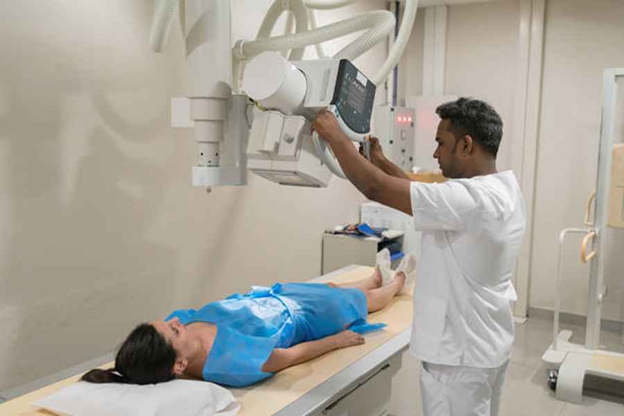
How is the scan done?
The technician (radiologist) to conduct your chest X-ray will collect your request form to ensure that your biodata, the reason for the X-ray (provisional diagnosis or indication), and any available clinical data that your physician may provide are accurate.
This initial documentation is important to avoid a mix-up of your results with those of other clients.
Clinical data is important because, at the end of the procedure, the technician conducting the X-ray will provide their interpretation of the result before it is sent to your doctor.
- You will be conveyed to a changing area where you are to change into a medical gown, remove pieces of jewelry and glasses, and keep all your personal belongings.
- You will then move to the X-ray room, which is a specialized room with thick doors and very little illumination.
- Here resides the X-ray machine, a large movable metal device with a camera at one end.
- A newer, more portable X-ray machine exits that can be taken to the patient’s bedside to conduct a test.
- There are lead coverings provided, which may be applied to your genital area to absorb any X-rays that may escape, affecting the testes in males or the ovaries in females.
- Eating meals, snacks, and drinking fluids would have to be suspended until the procedure was over.
- The technician will place you in position, in front or behind the X-ray camera, depending on the view required (front to back or back to front).
- This part requires you to take off your clothes from the waist up. You will be asked to stand straight and take a deep breath, then hold.
- This is done to prevent movement, which can distort the final image obtained after printing.
- Metal plates will be placed in front of and behind you; these contain the film that the final image obtained will be cast on.
- A painless beam of yellow light will be directed at your chest or back for less than a second, and the procedure is over.
- This will be followed by the printing of the result.
What should you do after the scan?
After the procedure is over, you will change back into your clothes and wait some minutes for the result to be printed, reported, and given to you to take to your doctor, who will make the final diagnosis based on your clinical data and findings from the chest X-ray.
Possible scan outcomes
Undergoing an X-ray procedure temporarily exposes an individual to radiation that can be harmful.
When conducted by a professional and the radiation is applied at a clinical dose, the risk of harm is greatly reduced.
Some measures can be employed to ensure safety when undergoing an X-ray, such as:
- Only go for an X-ray when a doctor requests the test.
- Your doctor will direct you to reliable technicians or units to conduct the procedure. This is particularly important because, due to the inexpensive nature of the X-ray machine, it can be handled by quacks.
- Inform your doctor about pregnancy if it is present. Although the procedure is essentially harmless to you, it may harm an unborn baby.
- Garments and other protective items that can absorb radiation are provided in the X-ray room.
The amount of radiation directed to the body during an X-ray is very minimal and cannot cause any significant problems.
You may, however, experience some discomfort from the ambient temperature of the room in which the X-ray is conducted.
There is a risk of aggravating pain from injuries to chest structures.
These, if experienced, would be temporary.
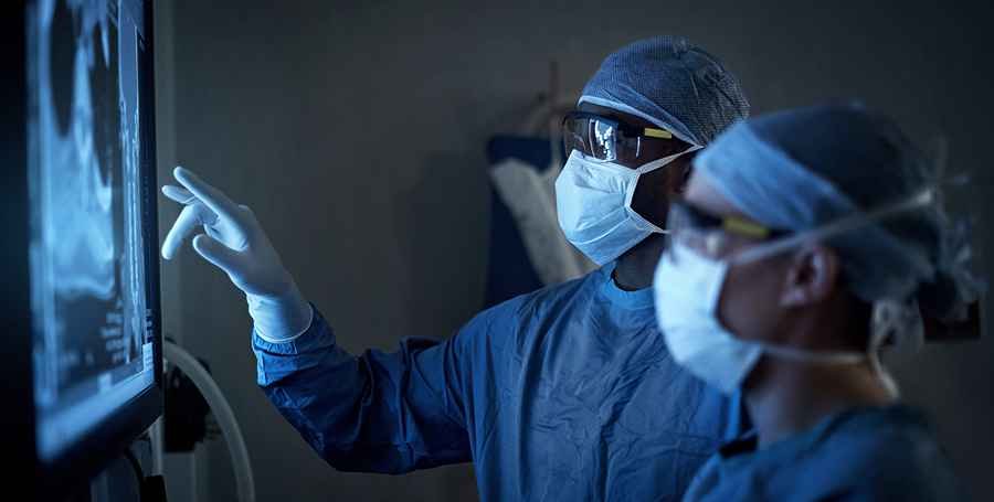
Interpretation of scan results
The X-ray technician and the doctor who requested the X-ray will give an independent interpretation of the result, but all will be guided by the clinical data and the findings on the X-ray film.
The result obtained from your X-ray may show no abnormality; in such a case, your doctor may consider other tests such as a CT scan or MRI, a blood test, or a biopsy, depending on the working diagnosis.
For example, some cancers in their early stages may not show any findings on chest X-rays.
On the other hand, the result may show abnormalities to support the clinical diagnosis or may show other abnormalities that have not manifested as symptoms yet.
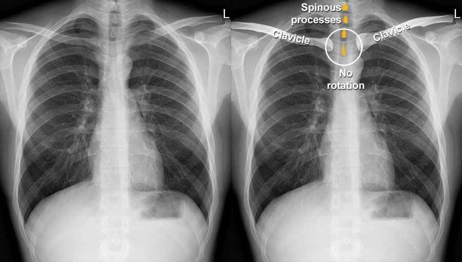
When the beam of X-rays is passed, they penetrate the body, and different physical elements or matter within the body (air, liquid, or solid) will absorb the radiation at different intensities depending on the densities of the elements or matter.
The higher the density of the matter, the greater the absorption of the X-rays, and the whiter its image will appear on the film.
- Radio-Lucent: Black or faintly grey appearance due to no or little absorption of the X-rays, respectively
- Radio-opaque: thick gray to white appearance due to absorption of X-rays
Note the following:
- Air does not absorb any of the X-ray waves; therefore, on the printing, it appears black.
- Fluid within the lungs, chest cavity, or space surrounding the heart (pericardium) will appear as a continuous white image on the X-ray film.
- Soft tissue, such as fat, blood vessels, and lymph nodes, will appear greyish-white.
- The bone will appear white because it has a high absorption of X-rays.
- Metals, plastics, or other non-biological elements will appear completely white, and often their outline or shape will be well-defined.
- The appearance of tumors on the chest X-ray depends on the composition of the tumor. Therefore, it may take any form; however, most tend to be whitish.
Possible diagnosis from a chest X-ray
1. Heart
- Hypertension
- Heart failure
- Infection involving the heart (carditis)
- Fluid collection surrounding the heart (pericardial effusion)
2. Lungs
- Tuberculosis
- Pneumonia
- Chronic obstructive pulmonary disease (COPD)
- Cystic fibrosis
- Pneumothorax (collection of air in the space surrounding the lungs and the chest wall)
- Lung cancers
3. Bones
- Rib fracture
- Clavicular fracture
- Spinal deformities such as Scoliosis (S-shape bending of the spine) and tuberculosis of the spine (Pott’s disease)
4. Blood vessels
- Suspected pulmonary Embolism
Conclusion
The diagnostic benefit of X-rays outweighs the risk of exposure to radiation during the procedure.
Your doctor would request an X-ray only when it was indicated based on your symptoms.
Feel free to talk to your doctor about any further concerns you may have about the procedure.
X-rays are safe and constantly required by physicians to aid diagnosis and monitor the progress of therapy.
If pregnant, inform your doctor when an X-ray is requested.
Importantly, adhering to all safety instructions during the procedure reduces the risk of excessive radiation exposure.