Even as a third-year medical student, pronouncing echocardiography stressed me out.
I mean, not another ‘raphy‘ or ‘logy’ to learn, but as we are about to see, echocardiography, or echo for short, is an important test often needed by the doctor to properly treat you.
This is especially true if he suspects you have a heart-related condition or are preparing for surgery.
Echocardiography is a test used to visualize the heart and its structures.
It uses sound waves to produce a live image of the heart called an echocardiogram.
Some of the heart structures it visualizes include
- Heart sac
- Its Valves
- Major blood vessels
- Flow and pressure of blood in the heart
- Heart rhythm
Reasons you might request the scan
Some of the reasons for an echocardiogram include:
- An irregular heartbeat or pulse rate picked up during a routine examination
- Persistent weakness and tiredness, especially after little activity
- Persistent chest pain, especially in the center of your chest between your ribs
- Shortness of breath
- If you’re going for major surgery and you are above 40, your doctor may also ask for one
Sometimes an echocardiogram can be requested in infants if there is some suspicion of congenital anomalies; some anomalies can go undetected until later in life.
Types of echocardiography
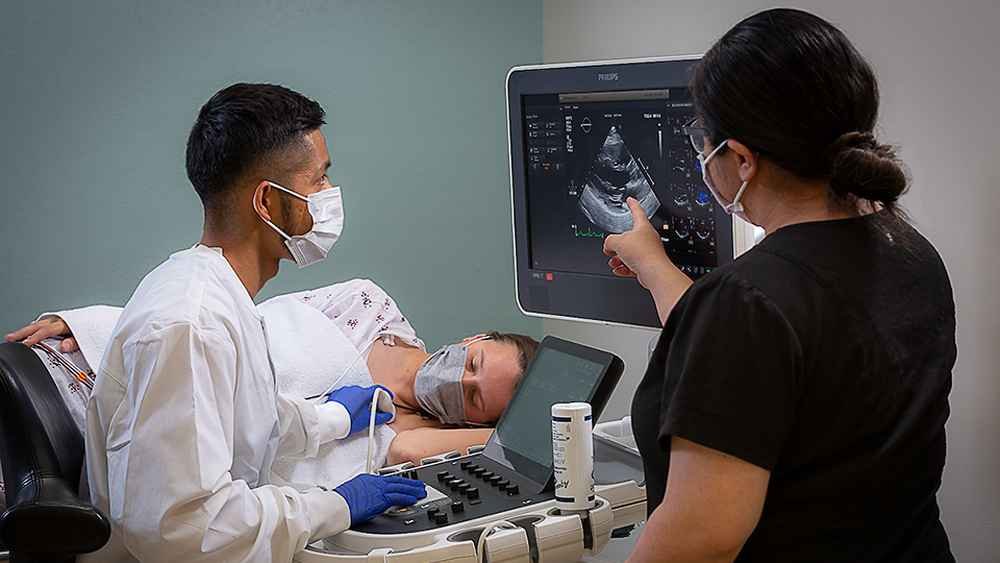
- Thoracic echocardiography: This is also referred to as standard echocardiogram, Electrodes are placed on your chest while a transducer probe is moved up and down your chest to visualize the heart
- Oesophageal echocardiography: This is a special type of invasive echocardiography where the probe is put down your throat until it gets close to your heart, from where an image is generated
- Stress echocardiography: This is similar to thoracic echocardiography, just that it is done after about 10–15 minutes of exercise
What should you do before the scan?
Usually, going for an echocardiogram needs no special preparation.
The most common type of echocardiography, transthoracic, is painless and involves placing a transducer over your chest, which sends sound waves to your heart and uses these to create an image.
This is completely painless and stress-free.
A certain type of echocardiography known as an esophageal cardiograph involves placing a transducer through your throat down to just behind your heart.
For this type of echocardiograph, you may be asked not to eat to prevent vomiting.
You would also need assistance driving or commuting home due to the sedatives given during this procedure.
How is the scan done?
- You’d be asked to undress from the waist up
- You then lie down on an examination bed
- Electrodes are placed on key points on your chest
- A transducer with some gel on it is then moved up and down your chest to visualize your heart
- You may be asked to make some postural movements for a clearer view
For Transesophageal echocardiography, however,
- An anesthetic spray or gel will be placed in your throat
- You would be given some sedatives to relax
- The tube with the transducer is moved up and down your throat to get clear images of the heart
What should you do after the scan?
If you did a standard echocardiogram, you can return to your normal activity immediately.
With a transesophageal echocardiogram, you may be asked to wait for some minutes for observation.
Also, you may need some assistance commuting home until the effects of the sedatives wear off, which usually takes a few hours.
Possible test outcomes
The echocardiogram would help rule out or confirm the suspicions of your doctor.
Possible abnormal findings include any of the following:
- Enlarged heart muscles
- Problems with the heart’s pumping mechanism
- Weak heart valves
- Constricted or enlarged heart blood vessels
- Presence of blood clots in the heart
Interpreting your results
Your results would come out as a black-and-white image that outlines your heart and its structures, usually accompanied by notes from the radiologist.
The results would be interpreted by your doctor, who may refer you to a cardiologist for further management after a complete assessment
Conclusion
So yes, echocardiography is essentially a test that helps view your heart and its structures as well as determine the integrity of its function.
It is usually painless and requires little preparation, except for the transesophageal one.
Have you had echocardiography done before? What was your experience like?
We would love to know; share them in the comment section.