In the realm of medical imaging, computed tomography pulmonary angiography (CTPA) stands as a powerful diagnostic tool, playing a crucial role in the assessment of pulmonary vascular conditions.
This blog post aims to unravel the intricacies of CTPA, shedding light on its applications, procedures, and significance in the diagnosis and management of pulmonary embolism and other vascular disorders.
What is the CTPA?
A CTPA scan, or computed tomography pulmonary angiography, is a specialized X-ray technique that uses a contrast dye to visualize the blood vessels in your lungs.
Think of it like a detailed map of your lung’s circulatory system, highlighting any blockages or abnormalities.
It combines the principles of computed tomography (CT) with contrast-enhanced visualization of the pulmonary arteries.
This imaging modality is designed to provide detailed, cross-sectional images of the pulmonary vasculature, aiding in the diagnosis of conditions affecting blood flow to the lungs.
Reasons why your doctor might request a CTPA
1. Pulmonary embolism (PE)
CTPA is most commonly employed in the evaluation of suspected pulmonary embolism.
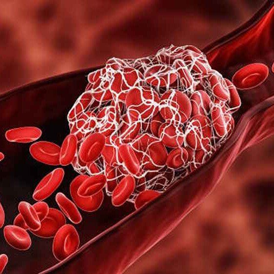
Pulmonary embolism occurs when blood clots, typically originating in the deep veins of the legs, travel to the lungs, potentially causing life-threatening complications.
CTPA allows for the visualization of these clots within the pulmonary arteries, aiding in prompt and accurate diagnosis.
2. Evaluation of pulmonary artery abnormalities
CTPA can be used to assess the pulmonary arteries for abnormalities such as aneurysms, stenosis, or congenital malformations.
This is particularly relevant in cases where patients present with symptoms or risk factors suggestive of vascular disorders.
3. Assessment of pulmonary hypertension
In cases of suspected pulmonary hypertension, CTPA can provide valuable insights into the pulmonary vasculature, helping to identify potential causes and contributing factors.
4. Preoperative planning
CTPA is utilized in preoperative planning for certain cardiothoracic procedures, providing detailed anatomical information that aids surgeons in preparing for interventions.
How the CTPA is done

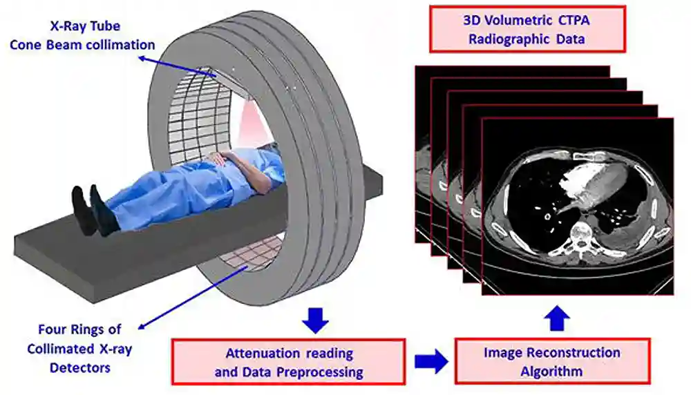
A CTPA scan is typically an outpatient procedure that takes about 15–30 minutes. Here’s a quick rundown of what to expect:
- Preparation: You’ll lie on a scanning table and may be given an intravenous (IV) line to inject the contrast dye.
- Contrast injection: Before the scan, a contrast dye containing iodine is injected into a vein, usually in the arm. This contrast material enhances the visibility of blood vessels on the CT images.
- Image acquisition: The patient is positioned on the CT scanner table, and the scanner rotates around the body, capturing detailed cross-sectional images of the pulmonary arteries. You may need to hold your breath for a few seconds at certain points to ensure clear pictures.
- Image reconstruction: The acquired images are processed by a computer to create detailed three-dimensional reconstructions, allowing clinicians to visualize the pulmonary vasculature in fine detail.
- Recovery: After the scan, you can usually go home right away. Drink plenty of fluids to help flush the contrast dye out of your system.
- Results: Your doctor will analyze the CTPA scan images and discuss the results with you. Depending on the findings, they may recommend further tests, medication, or other treatment options.
Risks associated with CTPA
While CTPA is generally considered safe, it involves exposure to ionizing radiation and the use of contrast material.
- Allergic reaction to the contrast dye: rare but can range from a mild rash to life-threatening anaphylaxis.
- Radiation exposure: While the amount of radiation is small, it’s important to discuss any concerns with your doctor, especially if you’re pregnant or have had multiple scans in a short time.
- Certain health conditions: It may not be suitable for certain patient populations, such as pregnant individuals or individuals with impaired kidney function.
The decision to undergo CTPA should be made in consultation with healthcare providers, weighing the benefits against potential risks.
Obisesan Damola
Damola is a medical doctor who has worked in the Nigerian healthcare industry for a little over 3 years in a number of primary, secondary, and tertiary hospitals. He is interested in and writes about how technology is helping to shape the healthcare industry. He graduated from the College of Medicine, University of Ibadan, the foremost medical training institution in Nigeria.


